


When it comes to taking care of your heart, choosing between ECG or Echo is really important. Electrocardiogram (ECG) and Echocardiography (Echo) are two important diagnostic tools that doctors use to check the performance of heart. ECG records the electrical activity of heart while echocardiography records structural images of heart. Understanding these tests and when to get them is essential for your heart’s health.
What is Electrocardiogram (ECG)?
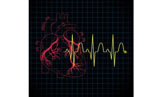
An Electrocardiogram, commonly known as an ECG or EKG (from the German term “Elektrokardiogramm”), is a non-invasive diagnostic tool used to record the electrical activity of the heart over a period of time.

Types of ECG
ECGs are of several types, including:
- 12- lead Resting ECG: It is conducted while the patient is at rest. This type provides a baseline measurement of heart electrical activity.
- Stress ECG (Exercise ECG or Treadmill Test): This test is performed during physical exertion, often on a treadmill or stationary bike, to assess the heart’s response to physical stress or increased demand.
- Holter Monitor (Ambulatory ECG): A portable device continuously records heart activity over 24 to 48 hours, providing a detailed analysis of daily electrical rhythms of heart.
- Signal Averaged ECG (SAECG): It’s a type of ECG which records the average of QRS complex over a period of 10 minutes or at least 250 QRS complex average. This is done to check small changes that can’t be identified on simple 12-lead ECG.
- Event Monitor: It is similar to the Holter monitor but it is used for more extended monitoring which can extend up to several weeks.
- Cardiopulmonary Exercise Test (CPET): This test assesses how the heart and lungs respond to exercise. It helps in providing valuable information about overall cardiac and respiratory function.
How ECG Works?
- Electrodes are attached to the chest, arms, and legs to detect electrical signals.
- It captures electrical impulses during each heartbeat.
- Received signals are amplified and recorded as ECG tracing.
- Cardiologist interprets for irregularities in heart’s electrical activity.
Conditions requiring ECG
- Detecting Arrhythmias: ECG helps in identifying irregular heart rhythms, such as atrial fibrillation or bradycardia.
- Assessing Heart Attacks: It plays a crucial role in diagnosing myocardial infarctions (heart attacks) by detecting changes in the heart’s electrical activity.
- Assessment of Cardiac Function: ECG can provide information about the overall function of the heart’s electrical system.
- Cardiopulmonary Assessment: The Cardiopulmonary Exercise Test assesses how well the heart and lungs work together during physical activity.
ECG Procedure
- The patient typically lies down, and small electrode patches or leads are attached to specific locations on the chest, arms and legs.
- The machine records the heart’s electrical signals, producing a graphical representation on paper or electronically.
- The results are interpreted by a cardiologist or healthcare provider.
What is Echocardiogram (Echo)?
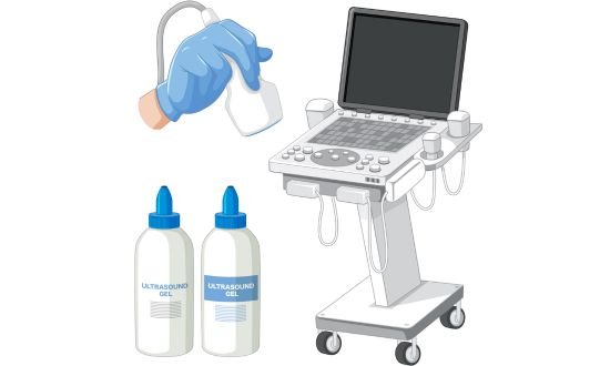
Echocardiography which is also called Echo, is a diagnostic imaging technique that uses high-frequency sound waves (ultrasound) to create detailed images of the heart’s structure and function.
Types of Echocardiogram
Echocardiograms come in various forms:
- Transthoracic Echocardiogram (TTE): It is the most common type. TTE involves placing a transducer on the chest to obtain images of the heart through the chest wall.
- Transesophageal Echocardiogram (TEE): It is more invasive but highly detailed procedure. TEE involves inserting a probe through the esophagus to get closer views of the heart.
- Doppler Echocardiography: This type assesses blood flow through the heart’s chambers and blood vessels.
How Echo Works?
- Gel application for sound wave conduction, followed by transducer placement.
- High-frequency sound waves emitted towards the heart.
- Sound waves bounce back upon encountering heart structures.
- Real-time images of heart, including chambers, valves, and blood flow are formed.
- Cardiologist interprets images to assess the heart’s structure and function. This helps in identifying heart structure’s abnormalities.
Conditions Requiring Echo
Echo plays an important role in diagnosing various heart conditions. Most common indications for echocardiogram are:
- Assessing Structural Anomalies: Echo is excellent for detecting structural issues like heart valve problems, congenital heart defects and heart muscle abnormalities.
- Evaluating Cardiac Function: It assesses how well the heart pumps blood which helps in diagnosing conditions like heart failure.
- Monitoring Heart Disease: Echo is valuable for tracking the progression of heart diseases and evaluating the effectiveness of treatment.
Echo Procedure
- A gel is applied to the chest to improve sound wave conduction and a transducer is moved across the chest.
- High-frequency sound waves bounce off the heart structures, creating detailed images on a screen.
- A cardiologist interprets the images to assess the heart’s structure and function.
Differences Between ECG and Echo
| Aspect | ECG | Echo |
| Diagnostic Focus | Electrical Activity | Structural and Functional Imaging |
| Invasiveness | Non-invasive | Mostly Non-invasive (TEE is invasive) |
| Continuous Monitoring | No | No |
| Images Produced | Graphical ECG Traces | Detailed Ultrasound Images |
| Primary Use | Detect Arrhythmias and Heart Attacks | Assess Structural Anomalies and Function |
| Typical Setting | Office or Hospital | Office or Hospital |
Potential Side Effects
Both ECG and Echo are generally safe procedures with minimal risks. However, rare side effects may include skin irritation from ECG electrodes or discomfort during a TEE procedure. It’s essential to discuss any concerns with your healthcare provider before undergoing these tests.
Frequently Asked Questions abut Ecg or Echo
1. Are ECG and Echo the same?
No, they are used for different purposes. ECG records electrical activity, while Echo creates images of the heart’s structure and function.
2. Do these tests hurt?
No, ECG is painless, and Echo is generally discomfort-free. However, TEE may cause mild discomfort.
3. How long does each test take?
ECG takes a few minutes, while Echo may take 30 minutes to an hour, depending on the type.
4. Are ECG or Echo always accurate?
These tests provide valuable information but the accuracy of the result depends on the patient’s condition and the skill of the healthcare provider interpreting the results.
5. Are there any age restrictions for these tests?
No, ECG and Echo can be performed on patients of all ages, from infants to the elderly.
6. Can I eat or drink before these tests?
For a standard ECG, there are typically no dietary restrictions. However, your healthcare provider may provide specific instructions for more specialized tests or Echo procedures.
7. Is it necessary to remove clothing for these tests?
You may need to remove clothing to access the areas where electrodes or the transducer will be placed. However, you’ll be provided with a gown or draping for privacy.
8. Can anxiety affect ECG or heart rate results?
Anxiety can indeed influence the results of an ECG (Electrocardiogram) or heart rate readings.
Conclusion
It is very important to understand the differences between ECG and Echo in order to choose the best tool for diagnosis of your specific heart condition. ECG records electrical activity of heart which helps in diagnosing conditions like arrhythmias, heart blocks and myocardial infarction. Echocardiography helps in diagnosing structural abnormalities of heart by providing detailed imaging of heart structure. Your doctor will recommend you the suitable test as per your specific medical condition.
Disclaimer
Content on this site is written with thorough research and keeping in mind the latest guidelines. However, no content on this site should substitute professional consultation.